
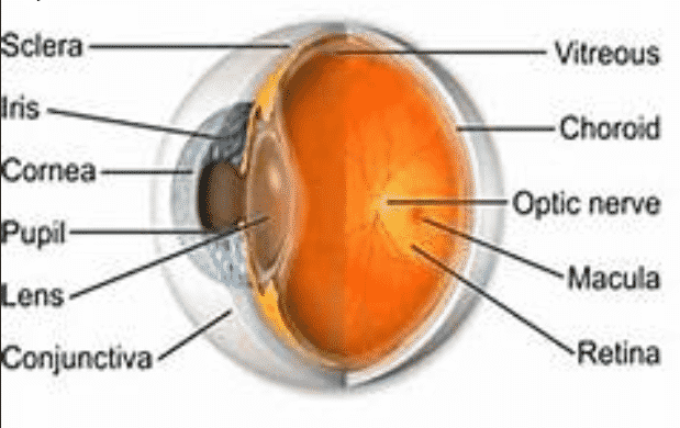
Health Editor’s Note: Without a functioning retina, sight is impossible. The retina lines the back of the eye and translates the light and images that come in through the front of the eye and translates an image into electrical neural impulses to the portion of the brain that interprets what is being seen. There are three types of photoreceptors in the retina. Rods which provide black and white vision and function in dim light, cones which interpret color and photoreceptor cells for responses to the brightness of light. When light hits the retina there is a waterfall of electrical and chemical events that activate nerve impulses which are sent to the visual centers of the brain through the all important optic nerve. You need a healthy retina to process light and to see clearly….Carol
NIH Scientists Combine Technologies to View the Retina in Unprecedented Detail
Technique enables direct imaging of neural tissue; could lead to earlier detection of diseases affecting eye tissue.
By combining two imaging modalities — adaptive optics and angiography — investigators at the National Eye Institute (NEI) can see live neurons, epithelial cells, and blood vessels deep in the eye’s light-sensing retina. Resolving these tissues and cells in the outermost region of the retina in such unprecedented detail promises to transform the detection and treatment of diseases such as age-related macular degeneration (AMD), a leading cause of blindness among the elderly. NEI is part of the National Institutes of Health, and the paper was published online in Communications Biology.
“For studying diseases, there’s no substitute for watching live cells interact. However, conventional technologies are limited in their ability to show such detail,” said the paper’s lead author, Johnny Tam, Ph.D., Stadtman Investigator in the Clinical and Translational Imaging Unit at NEI.
Biopsied and postmortem tissues are commonly used to study disease at the cellular level, but they are less than ideal for watching subtle changes that occur as a disease progresses over time. Technologies for noninvasively imaging retinal tissues are hampered by distortions to light as it passes through the cornea, lens, and the gel-like vitreous in the center of the eye.
..
 and the passage of fluorescent indocyanine green dye through the same vessels.")
Videos show multimodal technique using adaptive optics and angiography to simultaneously see blood cells flowing through the smallest blood vessels in the eye (left) and the passage of fluorescent indocyanine green dye through the same vessels.
Tam and his team turned to adaptive optics to address this distortion problem. The technique improves the resolution of optical systems by using deformable mirrors and computer-driven algorithms to compensate for light distortions. Widely utilized in large ground-based space telescopes to correct distortions to light traveling through the atmosphere, use of adaptive optics in ophthalmology began in the mid-1990s.
The NEI researchers combined adaptive optics with indocyanine green angiography, an imaging technique commonly used in eye clinics that uses an injectable dye and cameras to show vessel structures and the movement of fluid within those structures. In an observational study involving 23 healthy subjects, the researchers found that the multimodal approach enabled them to see for the first time a complex unit of cells and tissues that interact in the outermost region of the retina. The unit includes light-detecting photoreceptors, retinal pigment epithelial cells, which nourish the photoreceptors, and the surrounding choriocapillaris, capillaries that supply the outermost region of the retina with blood.
A range of diseases, including AMD, Alzheimer’s, and atherosclerosis (hardening and narrowing of the arteries), disrupt the outermost region of the retina. The ability to visualize live retinal cells and tissues may shed new light on these conditions and could help doctors identify early signs of disease before a person has symptoms, when the disease may be more likely to respond to treatment.
The investigators tested the multimodal imaging technique on a patient with retinitis pigmentosa, a neurodegenerative disease of the retina, and discovered well-preserved RPE and blood vessels in areas of the retina where photoreceptors had died.
“In the past, we have not been able to reliably assess the status of photoreceptors alongside RPE cells and choriocapillaris in the eye,” Tam said. “Revealing which tissue layers are affected in different stages of diseases – neurons, epithelial cells, or blood vessels – is a critical first step for developing and evaluating targeted treatments for disease.”
The study was funded by the Intramural Research Program at the NEI.
NEI leads the federal government’s research on the visual system and eye diseases. NEI supports basic and clinical science programs to develop sight-saving treatments and address special needs of people with vision loss. For more information, visit https://www.nei.nih.gov.
About the National Institutes of Health (NIH): NIH, the nation’s medical research agency, includes 27 Institutes and Centers and is a component of the U.S. Department of Health and Human Services. NIH is the primary federal agency conducting and supporting basic, clinical, and translational medical research, and is investigating the causes, treatments, and cures for both common and rare diseases. For more information about NIH and its programs, visit www.nih.gov.
NIH…Turning Discovery Into Health®
Reference
Jung HW, Liu T, Liu J, Huryn LA, Tam J. Combining multimodal adaptive optics imaging and angiography improves visualization of human eyes with cellular-level resolution. Published online Nov. 14 Communications Biology. DOI: 10.1038/s42003-018-0190-8

Carol graduated from Riverside White Cross School of Nursing in Columbus, Ohio and received her diploma as a registered nurse. She attended Bowling Green State University where she received a Bachelor of Arts Degree in History and Literature. She attended the University of Toledo, College of Nursing, and received a Master’s of Nursing Science Degree as an Educator.
She has traveled extensively, is a photographer, and writes on medical issues. Carol has three children RJ, Katherine, and Stephen – one daughter-in-law; Katie – two granddaughters; Isabella Marianna and Zoe Olivia – and one grandson, Alexander Paul. She also shares her life with her husband Gordon Duff, many cats, and two rescues.
ATTENTION READERS
We See The World From All Sides and Want YOU To Be Fully InformedIn fact, intentional disinformation is a disgraceful scourge in media today. So to assuage any possible errant incorrect information posted herein, we strongly encourage you to seek corroboration from other non-VT sources before forming an educated opinion.
About VT - Policies & Disclosures - Comment Policy


